Copyright Interesting Engineering

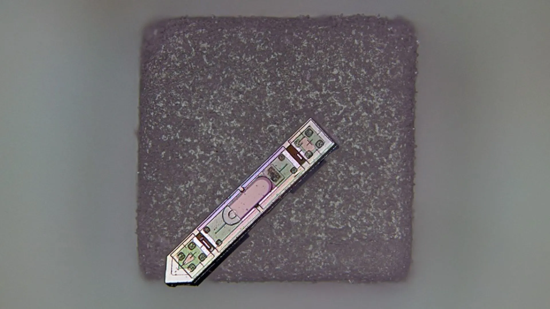
Cornell researchers have built a neural implant so small it can balance on a grain of salt, yet powerful enough to wirelessly record brain activity in a living animal for more than a year. The device, called a microscale optoelectronic tetherless electrode (MOTE), represents a major step toward long-term, minimally invasive neural monitoring and bio-integrated sensing. The project was co-led by Alyosha Molnar, the Ilda and Charles Lee Professor in Cornell’s School of Electrical and Computer Engineering, and Sunwoo Lee, an assistant professor at Nanyang Technological University who began developing the technology while working in Molnar’s lab. The implant measures about 300 microns long and 70 microns wide. Powered by harmless red and infrared laser beams that pass through brain tissue, the MOTE transmits data back through tiny pulses of infrared light that encode the brain’s electrical signals. A semiconductor diode made of aluminum gallium arsenide captures light energy to power the circuit and emits light to send data. It also includes a low-noise amplifier and optical encoder built with the same semiconductor technology found in everyday microchips. “As far as we know, this is the smallest neural implant that will measure electrical activity in the brain and then report it out wirelessly,” Molnar said. “By using pulse position modulation for the code – the same code used in optical communications for satellites, for example – we can use very, very little power to communicate and still successfully get the data back out optically.” Long-term tests in mice The researchers first tested the MOTE in cell cultures before implanting it into mice’s barrel cortex, the part of the brain that processes sensory information from whiskers. Over the course of a year, the implant consistently recorded both sharp neuron spikes and broader synaptic activity while the mice remained healthy and active. Molnar said the team wanted to design a system that avoided irritating brain tissue. “One of the motivations for doing this is that traditional electrodes and optical fibers can irritate the brain,” he said. “The tissue moves around the implant and can trigger an immune response. Our goal was to make the device small enough to minimize that disruption while still capturing brain activity faster than imaging systems, and without the need to genetically modify the neurons for imaging.” New possibilities for neuroscience The MOTE’s semiconductor composition could allow researchers to collect brain activity data even during MRI scans, something not feasible with current implants. The technology may also be adapted for use in the spinal cord or integrated into artificial skull plates for continuous monitoring. Molnar first conceived the idea in 2001, but development picked up about a decade ago when he began collaborating with members of Cornell Neurotech, a joint effort between the College of Arts and Sciences and Cornell Engineering. The study is published in the journal Nature Electronics.


