Copyright forbes
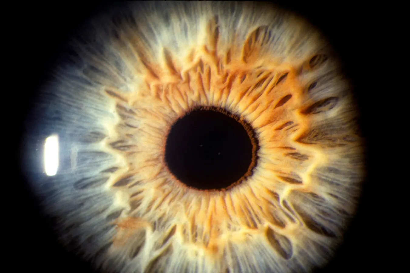
UNDATED PHOTO: A human eye is seen closeup, showing the iris and pupil. (Photo by Joe McNally/Getty Images) Getty Images From clinic bottlenecks to home OCT and foundation models, a new wave of “oculomics” could help prevent millions from losing sight—if regulation, reimbursement, and equity keep pace. More than 2.2 billion people live with near- or distance-vision impairment, and about 1 billion of those cases were preventable or untreated—meaning millions are still losing sight that could be saved. In England’s NHS, ophthalmology is now the busiest outpatient specialty, and the pressure on appointments often translates into weeks of delay for patients. Age-related macular degeneration (AMD) is the leading cause of vision loss in older adults. It damages the macula—the part of the retina for sharp, central vision—so people struggle to read, drive, or recognize faces; “wet” AMD can worsen quickly (often treatable but visit-intensive), while “dry” AMD, including geographic atrophy, progresses more slowly but steadily. This feature grew out of my recent video interview with Prof. Pearse Keane (UCL/Moorfields), where we explored how AI is moving from code to clinic and what that means for patients now and over the next few years. Watch the interview here Why should an eye patient care? Because AI is making eye care faster, simpler, and closer to you. It can deliver a yes/no answer during a regular visit (fewer extra appointments), help your doctor spot changes earlier (treatment on time), and, in some cases, let you monitor from home (fewer injections or trips without risking vision). The goal isn’t to replace your specialist—it’s to get you the right care at the right moment with less hassle. Code Meets Clinic: OCT, Autonomous AI, and Reimbursement “The brutal truth is that people are losing sight because of delays,” says Prof. Pearse Keane. He helped launch the Moorfields–DeepMind program that showed an AI system could analyze OCT scans at specialist level along a real clinical pathway—turning a research milestone into a clinical blueprint. He later co-led INSIGHT, now the world’s largest ophthalmic bio-resource with 35+ million eye images linked to outcomes—built with governance, privacy, and public engagement from day one. MORE FOR YOU The clearest “code-to-clinic” proof today is autonomous AI for diabetic retinopathy screening. Michael D. Abràmoff, MD, PhD (University of Iowa; founder, Digital Diagnostics) led the first FDA De Novo-cleared autonomous medical AI in 2018. Crucially, it’s reimbursable in the U.S. via CPT 92229, enabling primary-care, point-of-care diagnosis. In plain English: autonomous AI makes the clinical call (and carries liability), so it can safely sit where the patient is; assistive AI supports a clinician who remains liable. In real programs, time from screening to specialist follow-up has dropped from 1–2 months to 3–5 days, and clinics have reached top national quality benchmarks—even as U.S. adoption sits near ~2% because local economics and operations lag the evidence. By the Numbers (today): 2018: first FDA De Novo for autonomous DR AI; 2021: CPT 92229 created for autonomous retinal imaging; 2024: De Novo for home OCT with AI to remotely monitor wet AMD between office visits. Together these milestones mark the shift from code → clinic—and increasingly, clinic → home. Population Scale: Singapore’s National Eye Screening Playbook When Assoc. Prof. Daniel Ting looks back at Singapore’s national program for diabetic retinopathy, he describes a formula that scales. First, referral pathways were co-designed by primary care and ophthalmology so patients flagged by AI flowed to the right level of care—cutting unnecessary tertiary referrals and keeping frontline clinicians onside. Second, a national tele-ophthalmology platform stitched together fundus cameras and electronic medical records, so an image taken in a community clinic could be uploaded, analyzed, and reported back in minutes. Third, Singapore adopted an “AI-first, expert-second” operating model: algorithms screen every image, and only uncertain or positive cases escalate to human graders. Keeping a nationwide system safe required more than intent. Ting points to the reporting standards he helped champion—STARD-AI and DECIDE-AI—as the guardrails that turn “promising” into proven clinical practice. In real terms, that means transparent dataset reporting, clear subgroup performance results, early in-clinic evaluations, and ongoing drift monitoring across devices and demographics—with recalibration before wider scale-up if performance slips. And while researchers cite AUC, Ting argues the next headline metric for health ministries isn’t a lab accuracy score at all—it’s pathway-level cost savings: evidence that AI-enabled screening shortens turnaround, improves referral precision, and reduces avoidable specialist visits across the journey. Inside Health Systems: Changes Patients Actually Feel At Johns Hopkins’ Wilmer Eye Institute, T. Y. “Alvin” Liu, MD focuses on changes patients notice. The first is clerical: ambient AI scribes are beginning to replace manual note-taking, so clinicians face people, not screens. The second is clinical: for neovascular AMD on as-needed (PRN) treatment, home OCT or remote retinal imaging paired with AI is compelling if it preserves the same vision with fewer visits or injections. Liu’s 90-day “AI-ready clinic” checklist is plumbing, not pixie dust—an HL7/FHIR-compatible EHR, a simple alert workflow, and disciplined change management—because most failures are human-factor failures, not algorithmic ones. To keep adoption fair, he tracks real-world outcomes by race and ethnicity during rollout. Therapies Meet AI Precision: Anti-VEGF, Home OCT, Gene/Cell Tongalp H. Tezel, MD (Columbia) treats wet AMD at the front lines and runs a lab advancing gene- and cell-based therapies. His near-term view is practical: use AI-integrated OCT to personalize anti-VEGF injection schedules (patients aren’t all the same), and pair home OCT with AI triage so clinics time visits and injections better. In early reports, home-monitoring programs avoided unnecessary injections in ~42% of cases and triggered earlier intervention in ~35%—a signal that smarter monitoring can cut burden without sacrificing vision. The evidence bar, he stresses, is clinical: phase-3 randomized trials must show superior vision outcomes, not just faster fluid detection. Looking 2–5 years out, he expects AI to help match the right patient to the right therapy—from RPE cell replacement to gene therapy—using OCT features such as the ellipsoid zone, GA size, and subretinal hyperreflective loci. Rule of care: AI detects change; the retina specialist decides. To keep this safe and equitable, training/validation must span diverse populations and OCT devices, with real-world error and subgroup performance monitored as adoption grows. Foundation & Multimodal Models: From Pretrain to Patient Safety “Foundation and multimodal models are opening new avenues,” says Alan Karthikesalingam, MD, PhD (Google DeepMind). “These include assisting physicians with conversational clinical reasoning, providing patients with clearer information, and enabling natural-language interaction with model predictions. Adapting these capabilities to high-risk clinical care—and assuring safety and generalization—remains active work.” In practice, that means guardrails by default: transparent documentation, routine drift monitoring, and defined update/rollback procedures. On the research front, RETFound—a foundation model trained on ~1.6 million retinal images and released for non-commercial research—shows why “pretrain once, adapt many times” matters: fewer labels to fine-tune, stronger robustness across devices and sites, and potential sensitivity to rare-disease signals smaller datasets miss. As health systems move beyond pilots, cross-hardware durability plus live performance surveillance will be baseline requirements. A Signal From the Future: Implants for Geographic Atrophy In October, BBC World covered a multicenter trial of a subretinal photovoltaic implant (PRIMA) at Moorfields and other European sites that restored meaningful reading in late-stage dry AMD (geographic atrophy). It isn’t standard care and requires careful selection and rehabilitation, but it shows the pipeline’s direction. As Pearse Keane notes, AI + OCT/home monitoring can help identify who benefits and when, opening the door to implants, gene therapies, and cell therapies—with safety and equity as guardrails. Roadmap: Who Does What (Now → Next) Patients & Families Ask for point-of-care screening. If you have diabetes, ask about autonomous AI for diabetic retinopathy; it can give a same-visit answer and speed follow-up. Explore home monitoring. For wet AMD, vetted home OCT programs may keep vision the same with fewer visits/injections. Clinicians & Health-System Leaders Build the plumbing. In ~90 days: HL7/FHIR EHR, simple alerts, and a pause/update/rollback plan. Measure what matters. Track time-to-treatment, completion rates, and avoidable referrals; audit by device and demographic subgroup (equity). Scientists & Builders Choose your lane. Architect for autonomous AI (system liability; point-of-care scale) or assistive AI (clinician-in-the-loop). Own the lifecycle. Use foundation models for robustness and define drift triggers to pause/update/rollback—and report them. Policy Makers & Investors Align incentives. Codes like CPT 92229 enabled use; next, make local ROI transparent so clinics see the benefit. Fund the backbone. National tele-ophthalmology platforms, device connectivity, and reporting capacity are infrastructure. The Bottom Line Today, real-world deployments show AI in eye care works where patients live: autonomous AI for diabetic retinopathy in primary care (with reimbursement), national tele-ophthalmology pipelines that move images and results in minutes, and home OCT programs beginning to reduce unnecessary visits and injections—while AI scribes return time to clinicians. Next (12–36 months), expect foundation-model robustness, routine drift monitoring, and predictive retina AI to better time interventions. On the horizon, new implants, gene therapies, and cell therapies should arrive not as stand-alone miracles, but as options matched by AI to the right patient at the right moment under strong clinical governance. The eye exam is becoming an AI exam—bringing specialist-level care to where people are, so treatment is earlier, safer, and more equitable. Let’s focus on a blueprint for saving sight at scale. Editorial StandardsReprints & Permissions