CLEVELAND, Ohio — If an International Space Station astronaut breaks her leg, there’s no ambulance service to the nearest X-ray machine more than 200 miles away on Earth.
NASA Glenn Research Center in Cleveland wants to remedy that by leading the search for the perfect small, portable X-ray machine to fly into space.
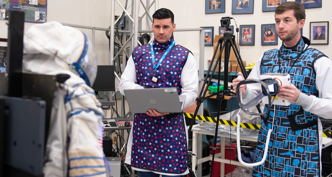
Three NE Ohio organizations — NASA, Cuyahoga Community College and University Hospitals — are collaborating to test portable, handheld X-ray systems that could be used on spacecraft for both medical and nonmedical uses.
The local effort is part of a larger NASA initiative to boost astronaut health and safety during long-duration spaceflight, where medical support is limited.
NASA is comparing three brands of portable, low-power X-ray devices to decide which is worthy of a space assignment. The chosen device must be easy for a non-technician to use, able to scan both human and inanimate objects, and produce hospital-quality X-rays.
The winning device will be selected by next year. It is scheduled to fly on the International Space Station in 2027 or 2028.
An X-ray machine on the International Space Station — or in future spacecraft bound for the moon or Mars —could be used to detect changes in the human body due to radiation or microgravity in space.
It also could examine parts of the spacecraft for damage, or check the health of a growing plant without uprooting it, said Cy Peverill, project task lead at NASA Glenn.
“Having the right tools on hand is crucial to ensure the survival of the crew,” Peverill said. “It costs a lot of money to send anything into space, so we need to show that these X-ray machines are worth the amount of room they would take in the crew module.”
Currently the International Space Station has an ultrasound machine but no X-ray, Peverill said. Ultrasound produces moving images, but is limited in its penetration depth and clarity.
NASA Glenn found the expertise needed for its X-ray evaluation project close to home, in Northeast Ohio.
Tri-C’s advanced radiography and dental hygiene labs taught the NASA team how to correctly use the X-ray devices.
UH afforded opportunities to test the mini X-ray devices on patients and compare their images with those produced by hospital-grade equipment.
“This has been a very multi-disciplinary project, and it was really cool that we were able to leverage all the expertise nearby,” said Chase Haddix, senior biomedical engineering research contractor working for Universities Space Research Association at NASA Glenn.
The third test site was NASA Glenn itself, where the devices took X-ray images of non-human objects such as rocks or electronic equipment.
A NASA Glenn lab specializing in tires for lunar rovers gave the X-ray project some intentionally damaged rover tires to experiment with.
“We’re very impressed so far with the ability of these low-powered X-ray devices to penetrate through space suits, metal objects and electronics,” Haddix said. ” … We were able to see inside (the tires) where some of the springs had bent, which could support a decision to switch out that tire before you go on a five-mile (trip) on the moon.”
NASA lands at Tri-C and UH
For a month, NASA staff practiced X-ray techniques at Tri-C’s advanced radiography lab, using human bones encased in clear plastic, called anatomical phantoms. The anatomical phantoms also are used by Tri-C’s students who are training to be radiographers who administer X-rays, said Elizabeth Gildone, program director for radiology and mammography at Tri-C Western Campus.
The NASA team also learned how to position X-ray equipment for the best image quality.
“There were all kinds of technical considerations they were learning from us, and we were learning what their needs were in the space environment,” Gildone said.
Students training to be radiographers watched the testing, learning from the NASA team. About 40 students graduate from the two-year program annually and go on to careers administering X-rays to aid in the treatment and diagnosis of disease, Gildone said.
Tri-C’s dental hygiene department at the Metropolitan Campus taught NASA investigators how to take a complete series of mouth X-rays using specialized manikins containing human bone, tissue and teeth from cadavers, said Anne Myatt, director of the college’s dental hygiene program.
“During this phase of their testing, it was important not to expose humans to ionizing radiation, but necessary to see how actual human dental structures would appear in the X-ray images,” Myatt said.
Faculty and students were asked to evaluate and give feedback about the X-ray images as part of the evaluation process.
NASA returned the favor by hosting Tri-C faculty, staff and students on a behind-the-scenes tour of NASA Glenn. “That was just a whole day of amazement and wonder,” recalled Tri-C’s Gildone.
The next step in the evaluation process was comparing the images made by portable X-ray devices with hospital equipment.
UH patients at the health system’s main campus currently are being offered the opportunity to have their X-rays taken twice — once with hospital equipment and once with NASA’s devices. UH physicians and NASA scientists will compare both sets of images, said David Jordan, chief medical physicist in radiology at UH.
Researchers aim to spend about a month collecting X-ray images of various body parts from a total of 40 UH patients, Jordan said.
“If they find, for example, that this equipment works well for wrists, ankles and forearms, but it’s not so useful for shoulders or chests, that’ll factor into their decisions for when and how they would pull it out during a mission,” Jordan said.
The project kicked off in June 2024, as NASA reviewed more than 200 X-ray machines for size, weight, image quality, ease of use and other qualities. The field was narrowed to three systems: MinXray, Remedi and Fujifilm.
The MinXray company, based in Northbrook, Illinois, is a key global supplier of portable, compact digital imaging equipment. The MinXray device, the largest of the three candidates, is about the size of a coffee machine. It recently took the first X-ray of a human hand in space during a Space X mission, the company said.
Korea-based Remedi is a global leader in medical technology innovation, according to the company website. A Remedi X-ray machine is scheduled to fly on a SpaceX mission in 2026.
Fujifilm, with world headquarters in Tokyo, is involved in a wide range of businesses healthcare, electronics and imaging.
Just like the faculty and students at Tri-C, UH staff were excited to work on a unique project that will help humans function better in space.
“There’s a certain amount of bragging rights to it all,” UH’s Jordan said. “We’re helping support the crew that’s going out there.”