Copyright Baton Rouge Advocate
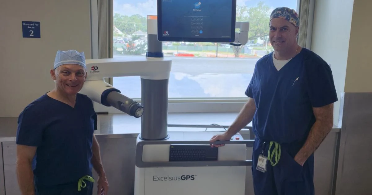
Louisiana doctors, Dr. Lawrence Haber and Dr. Ryan Farmer, have performed the first ever robotic-assisted pediatric spine surgery in Louisiana and the Gulf South. The duo at Ochsner Children's work with patients with varying degrees of scoliosis, a curving of the spine. About 2% to 3% of the U.S. population develop scoliosis — that's approximately 6 to 9 million people. The prevalence is higher in children with the primary diagnosis happening between 10 and 15 years old, according to the National Scoliosis Foundation. Up to 8% of patients with scoliosis may develop Scheuermann’s kyphosis, a structural spinal curve in the upper back that makes the top part of the spine rounded so it looks hunched over. The procedure to straighten the spine of a patient with Scheuermann's kyphosis involves surgically attaching various chords to vertebrae in the spine. Using the ExcelsiusGPS robotic system, Haber and Farmer were able to improve the accuracy of the placement of these chord. Although used in adults for two years, Farmer said the inclusion of the robot-assisted surgery for pediatric patients can lead the way to faster, safer recovery. Farmer lives in Lafayette, but travels across the state to see patients and perform procedures like the robotic-assisted surgeries as a pediatric orthopedic surgeon. Originally from Colorado, Farmer fell in love with the South and Louisiana when he got his master's degree in microbiology from the University of Louisiana at Lafayette. Even when he moved back to Colorado for medical school and postgraduate training, he came back to visit Louisiana a few times each year. He settled down in Louisiana with his wife in 2019 and has been at Ochsner Children's for two and a half years. What is scoliosis? What does it look like? Scoliosis is a 3D curvature of the spine. It's not only visible from the front and back sides of the body. There's a rotational piece of the puzzle that you can't necessarily see on an X-ray or in person. Sometimes we see it present as one side of the back looking longer than the other — that's a very visual cue that something is happening in a spine. Diagnostically, scoliosis is a curve in the spine greater than 10 degrees in a specific plane or direction. Curves in the coronal plane, facing the eyes, are all quite normal. Of the 2% to 3% of the population with scoliosis, 10% have a recommendation of surgery. What are the current treatments for children with spinal curves? Treatment is largely dependent on how big or how old a child is. The golden standard for care with scoliosis is monitoring. If the spinal curve is between 20 and 40 degrees, and there is some growth-time remain for the patient, we utilize a brace to prevent the curve from getting significantly worse. If the spinal curve is between 40 and 45 degrees, we recommend surgical intervention. In most studies about scoliosis, is a patient does not receive treatment, the curvature of their spine will get worse over time at a rate of about one degree per year. A 15-year-old with a 26-degree spinal curve might just need monitoring. But, if we let it go untreated or unmonitored, that patient may have a 60-degree curve in their spine at age 50. Once a spinal curve reaches 80 degrees, the spine starts to affect lung function making it difficult to walk long distances or do much activity. If we can catch these curves at an earlier time, at a young age, we feel that we can prevent these curves from reaching high-degree thresholds. Plus, kids heal significantly better than adults. In my mind, with scoliosis treatment, it's an important distinction between recommended surgeries and required surgeries. We are not treating an infection, a tumor or a fracture. We really have to do surgery to improve a patient's quality of life as they get older. We allow patients and their families some time to think about it as well. How have spinal treatments and technologies changed over the years? Spine fusion procedures introduced in the 1960s infused a Harrington rod, or metal rod, to the spine surgically in order to straighten spinal curves. Patients would be in body casts for an extended period of time to heal. In the 80s, spinal fusions moved to placing screws into the bony column of the spine within each individual vertebrae. Then, surgeons would attach that to the rod, a fixed object. Over the last 40 years, we have gained incredible knowledge of the normal balance of the spine. Implants have also improved in both their material science — like special threads to attach the screws and what the rods themselves are made out of. Past spinal fusions made the natural curve of the spine in the side place of the chest region straight, which causes huge balance problems. The spine should not be completely, 'ramrod' straight. What patient was the first to receive a robot-assisted surgery? Why was it an important step in pediatric care? The first patient that we treated was young man who had Scheuermann's — a specific type of disorder where a patient has a spinal curve in the frontal plane. There's a normal lean forward in the chest region where the spine curves naturally for balance. With Scheuermann's, a patient will have a more than significant curve in that area resulting in a fairly large hump on their back. When we do spinal fusions, we place the screws on each individual vertebrae (freehand). We use landmarks to find the bony column of the spine. We take and instrument and make a path along the spine. We are 93% accurate in placing those screws freehand in a safe fashion. With robotics assistance, we are 97% accurate. With the robotics, we are improving the accuracy of screw placement and reducing risks associated with the surgery. The robot itself has been used in adult spine surgeries for many years, but we can safely use the robot on our smaller patients as well. We used the robot-assisted arm in a patient who was 8, a young girl who had the body size of a 4-year-old. This patient had extremely small bony columns to place those screws, and having the ability to safely put those screws in was a huge win for both me and Dr. Haber. This robot, specifically, is used to hold a tube that instruments are then placed through. We are the ones examining X-rays and CT images to pick the best, and safest, pathway — the size, the length, how the implant goes into the body. That information that we decide and collect gets translated to the robot. The robot holds that tube for us so that we can place the screws and instruments in the appropriate positions. The robot simply acts as a third, unmovable hand in the operating room.