Lifesaving MRI tool offers sharper view of dangerous heart condition affecting millions worldwide
By Joshua Shavit
Copyright thebrighterside
Four-dimensional imaging is changing the way doctors diagnose and treat one of the most dangerous heart conditions. Scientists at the University of East Anglia have tested an advanced form of MRI that provides a sharper and more reliable view of aortic stenosis, a disease that affects millions of people worldwide. The results show that this technology can give doctors the clarity they need to treat patients sooner and with greater confidence.
Aortic stenosis is more common than many realize. In the United States, about five percent of adults over 65 live with it. In the United Kingdom, around 300,000 people are affected. The condition happens when the heart’s main outflow valve stiffens and narrows. That narrowing limits how much blood can leave the heart, straining the body and putting a patient’s life at risk.
Symptoms can be easy to dismiss at first. Shortness of breath, chest pain, fatigue, and dizzy spells may sound like simple aging or stress. But for someone with aortic stenosis, these signs can point to a valve on the verge of failure. Left untreated, the condition often worsens and can lead to heart failure or sudden death.
Doctors usually rely on ultrasound, also known as echocardiography, to measure blood flow and check the valve’s opening. While non-invasive and widely available, this approach has a flaw. Ultrasound sometimes underestimates how severe the narrowing really is. That can mean patients miss the critical window for surgery or other intervention.
Dr. Pankaj Garg, a consultant cardiologist at the Norfolk and Norwich University Hospital and lead researcher at Norwich Medical School, explained the challenge. “Aortic stenosis is a common yet dangerous heart condition,” he said. “At the moment, doctors use an ultrasound to diagnose the condition, but this can sometimes underestimate the severity of the disease, delaying vital treatment.”
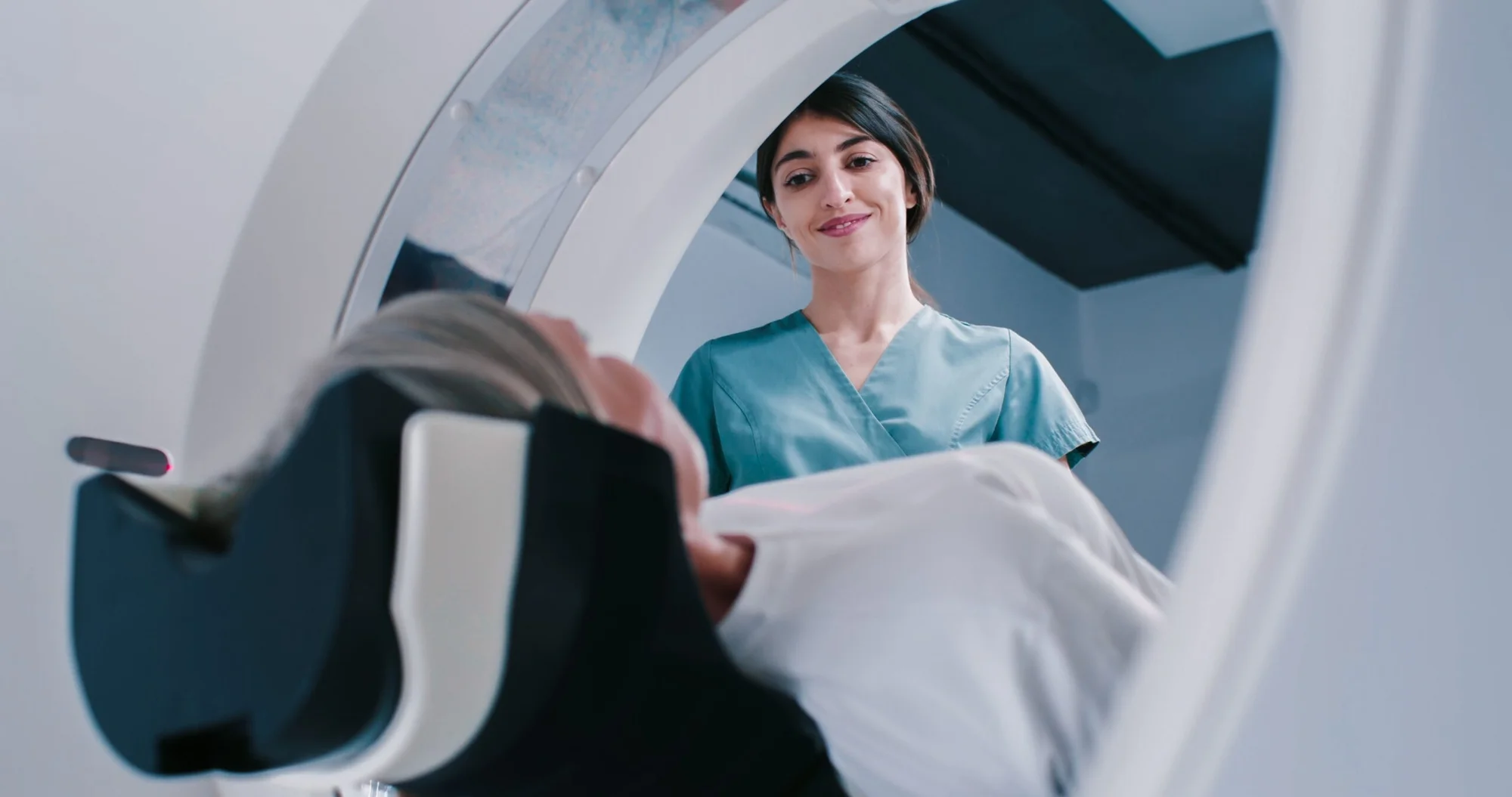
Researchers tested a different tool: four-dimensional flow MRI. Unlike standard scans, this technology doesn’t just create still images. It tracks blood moving in three directions, then adds time as the fourth dimension. That allows specialists to see blood flow in motion with remarkable detail.
“We wanted to see whether it could provide a more accurate and reliable diagnosis than a traditional ultrasound,” Dr. Garg said. The team studied 30 patients with confirmed cases of aortic stenosis. Each patient underwent both echocardiography and the advanced MRI.
The researchers compared the results, then followed patients for eight months to see which predictions matched their actual outcomes.
The results were striking. The MRI scans offered measurements that lined up more closely with what later happened in the clinic. By showing the true speed of blood passing through the narrowed valve, the scans gave a stronger signal about which patients needed timely surgery.
That difference could prove lifesaving. If doctors can spot severe narrowing earlier, they can schedule valve replacement before the heart weakens beyond repair. More accurate diagnosis also means fewer unnecessary operations for patients who may not yet need them.
Dr. Garg emphasized the potential. “4D flow MRI is an advanced heart imaging method that allows us to look at blood flow in three directions over time – the fourth dimension. We hope that this breakthrough will transform how cardiologists assess patients with aortic stenosis – leading to more timely interventions, fewer complications, and potentially thousands of lives saved in the UK alone.”
The findings build on earlier registry studies, such as the PREFER-CMR project, which compared ultrasound and MRI side by side. Those studies also found stronger agreement between MRI results and actual patient outcomes. What makes the latest work important is its focus on real-world clinical settings.
By following patients beyond the initial scans, the researchers could see how well each method predicted the need for intervention. The MRI scans proved to be the more consistent guide. This kind of validation matters because new medical tools often show promise in theory but struggle in practice.
The timing of treatment for aortic stenosis can be as critical as the surgery itself. Replace the valve too early, and a patient may face risks without much benefit. Wait too long, and the heart muscle may already be too damaged to recover.
That delicate balance makes reliable imaging essential. An MRI that captures the full flow of blood through the valve gives doctors a clearer map. With better data, they can choose the right moment to act.
Though promising, four-dimensional MRI is not yet routine. The technology requires advanced equipment, skilled operators, and more time than standard ultrasound. For many hospitals, those hurdles mean it cannot replace echocardiography overnight. But as the science grows and scanning systems become more efficient, adoption could spread. The research team hopes their findings will encourage wider testing in larger patient groups and across more healthcare systems.
Heart disease remains one of the world’s leading killers. Advances in imaging technology are reshaping how doctors detect and manage it. Four-dimensional flow MRI may soon give cardiologists a sharper lens to look inside the heart, helping them act before it is too late.
If widely adopted, the technology could change not just how patients are diagnosed but how lives are saved.
Research findings are available online in the journal Open Heart.
Note: The article above provided above by The Brighter Side of News.
Like these kind of feel good stories? Get The Brighter Side of News’ newsletter.