Copyright Interesting Engineering
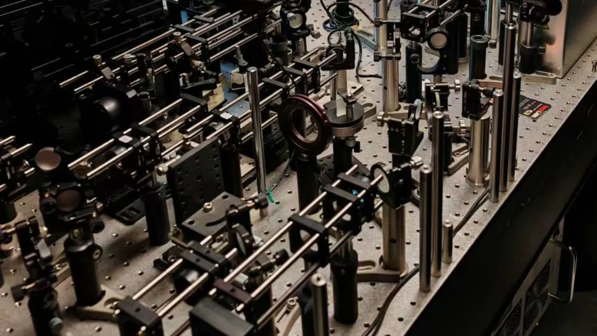
A research team from the School of Engineering at The Hong Kong University of Science and Technology (HKUST) has created the world’s first technology that captures high-resolution images of the brain of awake mice without anesthesia. This near non-invasive method allows scientists to observe brain tissue in a natural, fully functional state. The breakthrough could help researchers better understand how the human brain works in both healthy and diseased conditions, advancing neuroscience research in major ways. For decades, scientists have relied on imaging tools like MRI, EEG, CT, and PET scans to study the brain. These tools provide valuable data but lack the precision needed to capture the brain’s microscopic structures and rapid activity. Mice are often used as models for studying neurological disorders, cancers, and vaccines because of their genetic similarity to humans. However, anesthesia, commonly used during experiments, alters brain activity and blood circulation, affecting the accuracy of results. At the same time, the natural movement of awake mice tends to blur images, making it difficult to study the brain’s fine structures. New imaging system named MD-FSS The new technology, called Multiplexing Digital Focus Sensing and Shaping (MD-FSS), was developed by a team led by Professor Qu Jianan from HKUST’s Department of Electronic and Computer Engineering. MD-FSS builds on Qu’s earlier work, Analog Lock-in Phase Detection Focus Sensing and Shaping (ALPHA-FSS), which was published in Nature Biotechnology in 2022. While ALPHA-FSS achieved subcellular imaging accuracy, it was too slow to capture dynamic brain activity in awake animals. The skull’s thickness and density also pose challenges, as they scatter light and degrade image quality. These limitations have long prevented scientists from obtaining clear images of deep brain structures. To overcome this, the team designed MD-FSS to speed up how point spread function (PSF) measurements are made—the 3D image of a point-like object seen under a microscope. The system uses multiple weak laser beams along with one strong primary beam. Each beam carries unique spatial data and operates at different frequencies. Through digital phase demodulation, a method that isolates weak signals from noisy backgrounds, MD-FSS captures PSF data in less than 0.1 seconds, making it ten times faster than earlier techniques. The result is sharp, detailed images that track real-time brain activity. Sharper, faster, and deeper brain imaging By combining MD-FSS with multiphoton microscopy, the team created a new setup known as Adaptive Optics Three-photon Microscopy. This system can track immune cell movements, measure blood flow in tiny cerebral vessels, and monitor neuronal activity during thinking and sensing. It can even capture how brain cells and blood vessels interact. Qu said, “Such detailed, near noninvasive, and real-time observations in awake animals were previously impossible. With the rapid aberration-correction capability of this novel adaptive optics technology, high-quality imaging is now achievable without injuring the subject’s brain.” He added, “We can now capture the neuronal, glial, and vascular dynamics at subcellular resolution in their natural physiological state—free from the confounding effects of anesthesia. This breakthrough opens entirely new avenues for understanding brain function in both health and disease.” This imaging power is hundreds or even thousands of times sharper than EEG or CT scans. It enables researchers to see individual neurons, immune cells, and the smallest capillaries with remarkable clarity. Scalable design for the future of neuroscience The MD-FSS platform is built to scale. The current version uses eight laser beams but can be expanded to dozens or even hundreds. This scalability could make imaging even faster and allow scientists to explore larger areas of the brain as optical technologies evolve. Qu said, “Our latest work represents far more than an incremental improvement. We now have a versatile platform that can be scaled for faster imaging, expanded into larger brain regions, and integrated with functional assays.” He continued, “This will empower neuroscientists to investigate rapid brain events, complex network interactions, and disease progression in ways that were previously technically unattainable—opening the door to transformative discoveries in learning, memory, mental health, and neurological disorders.” The findings were published in the journal Nature Communications.