By Shrimansi Kaushik
Copyright deccanchronicle
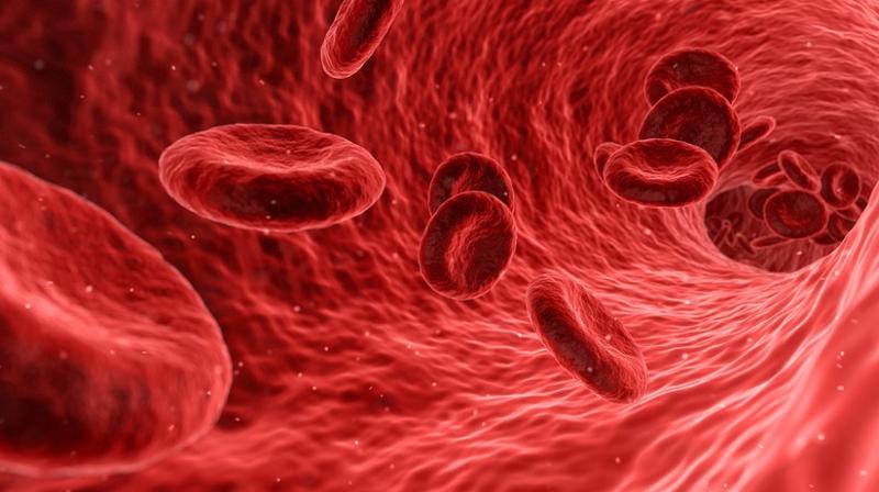
Hyderabad:Imagine a white blood cell chasing down a pathogen. To reach it, the cell pushes out flat, sheet-like protrusions that appear within microseconds, allowing it to “swim” toward its target. Scientists at the CSIR–Centre for Cellular and Molecular Biology (CCMB) have now discovered the molecular mechanism that enables such rapid shape-shifting. Their findings have been published in Nature Structural & Molecular Biology.A cell’s ability to change shape depends on its actin cytoskeleton — a dense, dynamic network of branched filaments beneath the cell membrane. Actin molecules can quickly assemble into new meshworks that push the membrane forward, forming protrusions. But how exactly do cells build these new scaffolds so efficiently?The CCMB team, led by Dr Saikat Chowdhury, has shown that a protein called SPIN90 plays a central role. They found that SPIN90 works as a dimer with another protein complex called Arp2/3. This initiates the growth of new actin filaments starting from the SPIN90-Arp2/3 complex, in two directions – always separated by about 150 degrees at the starting point.These new filaments serve as scaffolds for building a new branched actin meshwork. Thus, it creates a new protrusion, gives a new shape to the cell, or directs its motility.“SPIN90’s ability to build actin filaments in two directions may explain how cells rapidly create adaptable scaffolds,” said Dr Chowdhury. “This helps us understand how cells remodel themselves in health and disease, including cancer, immune disorders and wound healing,” he added.These findings let us understand the fundamental processes of cells. “We were able to see this mode of action of SPIN90 at nearly atomic resolution. This allowed us to understand the precise mechanism of actin filament formation from its earliest stages. It is the same mechanism that controls how all mammalian cells divide and function,” said Justus Francis, the study’s first author and a PhD student in Dr Saikat Chowdhury’s group at CCMB.”This breakthrough was made possible using CCMB’s advanced cryogenic electron microscope, provided by the Council of Scientific and Industrial Research (CSIR),” the researchers specified.