By Joseph Shavit
Copyright thebrighterside

Dementia has become one of the world’s most pressing health challenges. Millions of people live with the condition, and the global costs reach into the hundreds of billions each year. Doctors often rely on chronological age or blood-based markers to estimate risk, but these don’t always capture the underlying biology driving disease.
Now, researchers have discovered that the eye might provide an easier and more affordable way to forecast future brain decline.
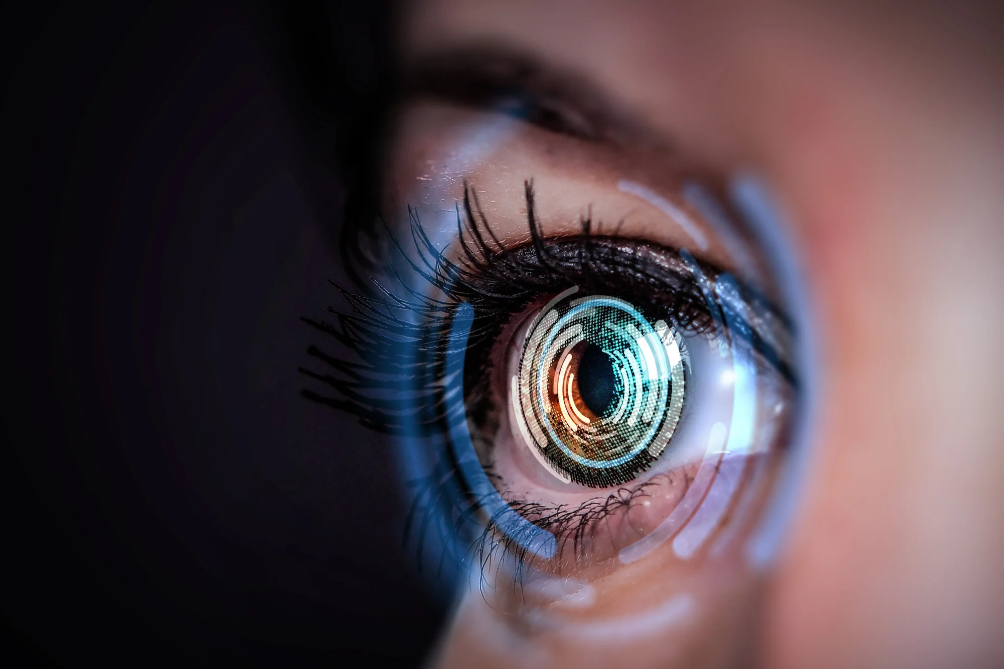
A team from the Yong Loo Lin School of Medicine at the National University of Singapore has created an artificial intelligence tool that analyzes standard retinal photographs to predict who is more likely to develop dementia or cognitive decline. The algorithm, named RetiPhenoAge, reads subtle patterns in the back of the eye to estimate biological aging and its link to brain health.
The study is the first of its kind in Singapore and one of the largest efforts to validate eye-based aging markers as predictors of dementia worldwide.
The retina is more than a sheet of tissue at the back of the eye. It develops from the same embryonic tissue as the brain and shares many of its features, including networks of small blood vessels. Because of this close relationship, scientists have long suspected that changes in the retina might reflect what is happening in the brain.
Professor Cheng Ching-Yu, Director of the Centre for Innovation and Precision Eye Health at NUS Medicine, explained, “With RetiPhenoAge, we are able to non-invasively estimate an individual’s biological age, offering valuable insights for both cognitive health management and broader ageing research.”
Unlike DNA methylation tests or brain scans, which require specialized facilities and come with high costs, retinal imaging is already widely used in eye clinics and polyclinics. That means a predictive tool built on these images could be deployed almost anywhere, including community health centers and mobile screening units.
The team built RetiPhenoAge using a deep-learning model trained on tens of thousands of retinal images from the UK Biobank, a major health study in the United Kingdom. The algorithm learned to estimate biological age based on a measure called PhenoAge, which combines nine blood-based biomarkers with a person’s actual age. Instead of relying on hand-picked features, the AI scanned the photographs for patterns linked to faster or slower aging.
Saliency maps—visualizations of what the algorithm focuses on—revealed that it paid close attention to blood vessels and the optic disc. This finding fits with biology, since small vessel disease and vascular aging play major roles in dementia.
The system was then tested in two large populations: the Singapore Memory, Ageing and Cognition Centre (MACC) cohort and the UK Biobank.
The MACC study followed 510 participants in Singapore over several years. At the beginning, they underwent eye photography, brain imaging, and cognitive assessments. Of these individuals, 155 developed measurable cognitive decline during follow-up.
Those with higher retinal ages as measured by RetiPhenoAge were far more likely to show decline or receive a dementia diagnosis. A three-point or greater increase in their Clinical Dementia Rating Sum of Boxes score was used to define cognitive decline. Each step up in retinal age meant a greater likelihood of decline, with hazard ratios of 1.34 for worsening cognition and 1.43 for new dementia cases over five years.
Professor Christopher Chen, Deputy Chair of the Healthy Longevity Translational Research Programme at NUS Medicine, said, “With dementia numbers rising globally, we urgently need tools that are both scalable and predictive. RetiPhenoAge could hold the key to community-level screening that is both effective and affordable.”
To ensure the tool worked beyond Southeast Asia, the researchers turned again to the UK Biobank. Among 33,495 participants with eye images and cognitive data, the algorithm once more predicted dementia risk. In this larger group, the hazard ratio for dementia was 1.25, confirming the method’s strength across different populations and ethnic backgrounds. The replication also extended the predictive window to 12 years, showing that retinal aging is a long-term signal, not just a short-term indicator.
The study went further by checking whether retinal aging tracked with measurable changes inside the brain. MRI scans revealed that people with higher retinal ages also had more white matter hyperintensities—signs of cerebral small vessel disease—as well as smaller hippocampal volumes, both of which are known markers of dementia.
Blood tests supported the link as well. Plasma proteomic analysis showed that proteins tied to inflammation, vascular aging, and neurodegeneration were elevated in those with older retinal ages. These findings suggest that the algorithm is not only picking up eye changes but also reflecting wider biological processes connected to aging and brain health.
Dr. Sim Ming Ann, Consultant at Ng Teng Fong General Hospital and co-first author, emphasized, “We hope that these findings will lead to improvements in care, which will help doctors identify people at risk of dementia before symptoms appear, which may lead to earlier interventions and improved patient outcomes.”
The global rise in dementia makes early detection more important than ever. Current estimates suggest that one in three seniors will experience some form of dementia, and numbers are expected to double in the coming decades. Tools like RetiPhenoAge could help identify those at highest risk long before symptoms appear, allowing for timely lifestyle changes, therapy, or monitoring.
Assistant Professor Tham Yih Chung, another co-first author, added, “We hope to validate this screening tool in larger and more diverse populations, and assess its impact in clinical settings to guide earlier treatment of dementia.”
While the results are promising, the researchers caution that the algorithm is not perfect. Not everyone with an older retinal age will develop dementia, and not every case of dementia can be predicted by eye scans. More studies in different global populations will be needed to refine the tool.
The team is already working on follow-up projects supported by a grant from the National Medical Research Council. These include testing retinal imaging in people with mild cognitive impairment and exploring how RetiPhenoAge might be used to track the effects of new therapies.
As Professor Chen noted, the goal is to make screening both simple and affordable, ideally as part of regular health check-ups. Since retinal cameras are already in wide use, the barrier to adoption is lower than for many other diagnostic technologies.
Research findings are available online in the journal Alzheimer’s & Dementia: The Journal of the Alzheimer’s Association.
Like these kind of feel good stories? Get The Brighter Side of News’ newsletter.


