Copyright NBC News
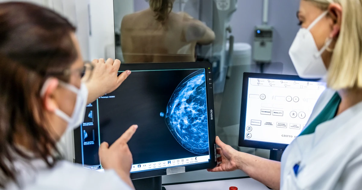
When a radiologist reviewed Deirdre Hall’s mammogram images last summer, everything seemed fine. There were no shadows or lumps or irregular patches that could signal cancer. The doctor gave it a second look for one reason: artificial intelligence software had drawn a circle around an area in the upper part of her left breast that it found suspicious. Because the AI software had put up that red flag, Hall, 55, got an order for an ultrasound that led to a biopsy. There were four cancerous tumors in the spot AI had identified. “This would have been completely missed without the AI,” said Dr. Sean Raj, chief medical officer and chief innovation officer at SimonMed Imaging in Tempe, Arizona, where Hall had her mammogram. Not only was Hall’s breast tissue dense, but the layers of tissue crisscrossed over each other in a particularly complicated pattern. “It camouflaged the cancer,” said Raj, a breast imaging specialist. “Even I could have missed it.” They caught her cancer at Stage 1, said Hall, who’s a respiratory therapist at a local hospital. “They didn’t find anything in the lymph nodes, which they were grateful for,” she said. “I’m so glad they caught it early.” When reading women’s routine mammograms, radiologists are increasingly augmenting their eyes with artificial intelligence. While many major medical centers have adopted the technology enthusiastically, some experts point to concerns, including a lack of studies in the U.S. showing that AI actually saves lives and does not needlessly raise concerns about benign growths. Experts train AI software by feeding it hundreds of thousands, or sometimes millions of mammogram images. Some of the images contain cancerous tumors, and, over time, the AI learns to distinguish the often subtle differences between malignant and benign tissue. Some AI programs, like the one used on Hall, identify a suspicious area. Others predict the chance that a woman will develop breast cancer. At the University of California, San Francisco, researchers are using AI to try to speed up the time from a mammogram to cancer diagnosis. In a study released this week, the radiologists used the technology to flag suspicious-looking mammograms so those patients could be seen more quickly. For patients with breast cancer, that AI triage cut the average time from mammogram to biopsy by 87%, from 73 days to nine days. The study was posted Tuesday to the preprint server MedRxiv. (Studies posted to preprint servers have not been peer-reviewed.) However, Dr. Sonja Hughes, vice president of community health at Susan G. Komen, a breast cancer organization, said more research is needed before AI is used as the standard of care. “We’re not there yet,” she said. “We don’t have enough research or enough data.” Dense breasts: Finding a snowball in a blizzard Mammograms have saved countless lives, but they’re imperfect. Dense breast tissue, which is a risk factor for developing cancer, makes mammograms harder to interpret. About 40% of U.S. women have dense breasts, according to the American Cancer Society. “It’s like trying to find a snowball in a blizzard,” said Dr. Otis Brawley, a professor of oncology and epidemiology at Johns Hopkins University. The Food and Drug Administration has authorized many AI programs for mammograms, with varying rates of accuracy. The AI software used on Hall’s mammogram, called Lunit, accurately identified cancers 88.6% of the time, according to a 2024 JAMA Oncology study of more than 8,800 women in Sweden who got mammograms. Another study published in Radiology noted that AI software caught cancers that were missed by two radiologists. However, in the Sweden study, AI gave a false positive 7% of the time, saying there might be a tumor when there wasn’t one. A false positive can trigger more testing and anxiety while waiting for results. With any mammogram, the chance of having a false positive result is about 10%, according to research. A doctor interprets the screening's results Major academic medical centers using AI in their imaging centers include the MD Anderson Cancer Center, the Mount Sinai Hospital in New York City, the Perelman Center for Advanced Medicine at the University of Pennsylvania, the Siteman Cancer Center at Barnes-Jewish Hospital, and MedStar Health. In all centers, the software is used along with, not instead of, a radiologist’s eyes, as FDA regulations require a doctor to interpret mammograms. Some breast imaging experts see advantages to this human-machine combination. “The nice thing about AI is that it doesn’t get tired,” said Dr. Lisa Abramson, associate professor of radiology at Mount Sinai. “It’s not going to replace the job or the expertise of radiologists, but I think it’s only going to enhance our ability to detect more and more breast cancers.” Brawley, the Johns Hopkins professor, said AI could help women who don’t have access to radiologists who specialize in breast imaging, and instead have their mammograms read by general radiologists. A study using RadNet’s software found that without AI, specialists correctly identified breast cancers 89% of the time, compared with 84% for generalists. With AI, the accuracy for both groups rose to about 93%. “It’s incredibly subjective when a human reads a mammogram,” Brawley said. “Maybe it’s going to reduce the disparities in how these things are read.” Does AI cost more? Typically, academic medical centers don’t charge patients extra for the use of AI software, and they can’t charge insurance companies for it, since there’s no billing code specifically for the AI, according toSusan G. Komen, a nonprofit breast cancer organization. SimonMed, which has centers in 11 states, and RadNet, which has centers in eight states, don’t charge for an initial layer of AI on mammograms, although patients are charged $40 and $50 respectively if they opt to have their images run through a second set of the technology. Drawbacks of AI Brawley worries that AI might be too good at its job. According to the American Cancer Society, it’s possible that mammograms flag some tumors that are technically cancerous, but not life-threatening. The patient then undergoes the physical, emotional, and financial toll of treating a tumor that was never going to hurt her. “It’scancer, but it’s not genetically programmed to grow, spread, or kill,” Brawley said. “I am worried that AI may help us find even more of these tumors that don’t need to be found.” Brawley pointed to the lack of data in the U.S. that shows AI actually saves women’s lives. Last month, researchers at the University of California, Los Angeles and University of California, Davis, announced a $16 million, two-year study at seven medical centers to take a deeper look at the technology. There are several other concerns about using AI in mammography. The technology isn’t perfect, and some worry that doctors could make mistakes if they become too dependent on it, according to an article last year in RadioGraphics. That’s why radiologists emphasize that AI is a tool, not a solution in itself. “It’s not going to replace the job or the expertise of radiologists,” said Abramson, the breast radiologist at Mount Sinai. “I think it’s only going to enhance our ability to detect more and more breast cancer.” Another concern is that if AI is trained mainly on breast images of white women, it could be less accurate for women of color, since genetic differences can make tumors look different. Hall, the Arizona patient, said she’s not necessarily a fan of AI in general — she says she finds the technology “creepy” — but she’s glad she paid $50 for the extra AI on her mammogram. “I don’t love all this AI stuff, but I definitely love this for me or anyone else in my position,” she said. “No matter how it was found, I’m glad it was found.” Guidance for mammograms Guidance from the United Services Preventive Services Task Force recommends women to get a mammogram every other year starting at age 40. According to American Cancer Society guidelines: Women 45 to 54 should get mammograms every year. Women 55 and older can switch to a mammogram every other year, or they can choose to continue yearly mammograms. Dr. Shanthi Sivendran, senior vice president at the American Cancer Society, offers guidance for more accurate breast cancer screening.