By Dr. Kamal Kant Kohli,Medha Baranwal
Copyright medicaldialogues
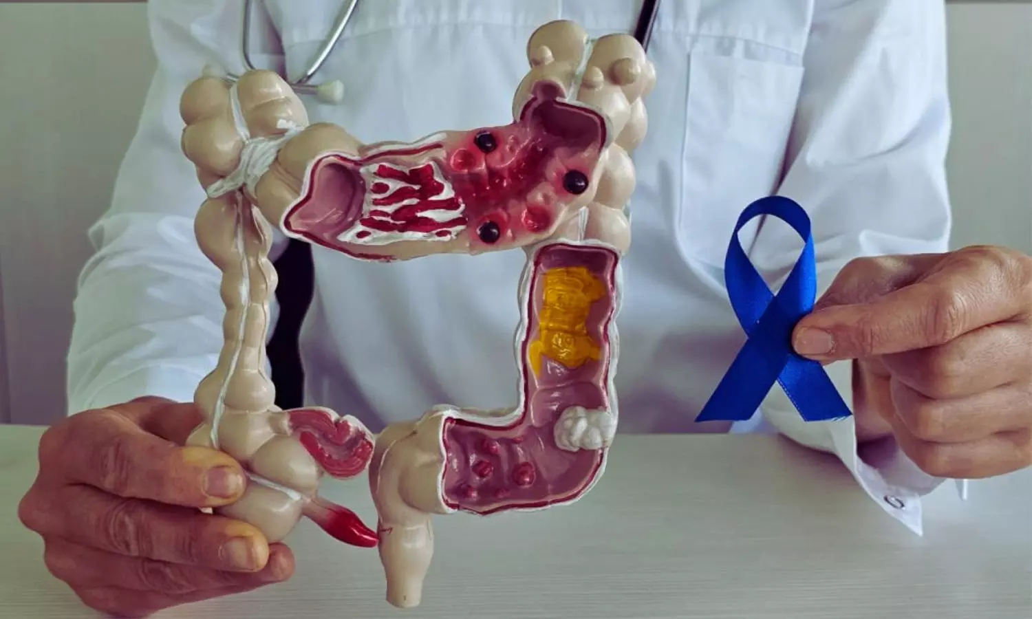
Poland: A recent prospective study published in the International Journal of Colorectal Disease has revealed that using PET/MRI instead of conventional imaging can substantially improve the diagnosis, treatment planning, and overall outcomes for patients with rectal cancer. The research shows that this hybrid imaging method provides superior accuracy in detecting lymph node involvement and tumor deposits, key factors in determining therapy.Rectal cancer remains a significant global health concern, with rising incidence and mortality, especially among adults under 50. The authors noted that in the United States alone, roughly 154,000 new colorectal cancer cases and nearly 53,000 deaths are expected in 2025. Accurate staging is therefore crucial to guide preoperative strategies and improve survival rates.The investigation, led by Dr. Rafał Maksim of the Maria Skłodowska-Curie Białystok Oncology Center in Poland, compared whole-body F-18 FDG PET/MRI with standard imaging techniques such as CT and MRI. Forty-two patients with histologically confirmed rectal cancer were enrolled. All participants first underwent conventional clinical staging and were scheduled for preoperative radiotherapy or radiochemotherapy. Before treatment began, they were restaged using PET/MRI to assess tumor size, lymph node status, and mesorectal tumor deposits. The study led to the following findings:PET/MRI showed the highest diagnostic accuracy for measuring tumor length, with an AUC of 0.73.Combining the maximum standardized uptake value (SUVmax) of lymph nodes with carcinoembryonic antigen (CEA) blood test results increased diagnostic performance to an AUC of 0.921.PET/MRI detected more lymph node involvement and mesorectal tumor deposits compared with conventional imaging.The hybrid scan changed the clinical stage in 64% of patients, including 11 upstaged, 16 downstaged, and 15 with no change.These findings led to modifications in treatment plans for about 26% of patients, affecting decisions such as surgical approach and the need for additional therapy.Hybrid PET/MRI combines detailed anatomical imaging from multiparametric MRI with metabolic data from PET scanning, offering a more comprehensive picture than either modality alone. While the study cohort was relatively small, the prospective design and the significant impact on staging emphasize the promise of this technology.The researchers emphasized the need for larger, multicenter prospective studies and long-term outcome tracking to confirm these findings and establish clear diagnostic thresholds.”Our results suggest that incorporating F-18 FDG PET/MRI into standard clinical pathways could become an important step toward more individualized and effective treatment for rectal cancer,” the authors concluded. Reference:Maksim, R., Buczyńska, A., Sidorkiewicz, I. et al. Standard of practice imaging vs. PET/MR: a comparative prospective study in rectal cancer staging. Int J Colorectal Dis 40, 198 (2025). https://doi.org/10.1007/s00384-025-04998-4